


Dental X-Rays
How do dental X-rays work?
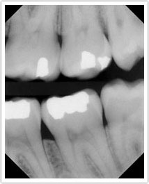
When X-rays pass through your mouth during a dental exam, more X-rays are absorbed by the denser parts (such as teeth and bone) than by soft tissues (such as cheeks and gums) before striking the film. This creates an image called a radiograph. Teeth appear lighter because fewer X-rays penetrate to reach the film. Tooth decay, infections and signs of gum disease, including changes in the bone and ligaments holding teeth in place, appear darker because of more X-ray penetration. Dental restorations (fillings and crowns) may appear lighter or darker, depending on the type of material used for the restoration. The interpretation of these radiographs allows the dentist to safely and accurately detect hidden abnormalities.
How often should radiographs be taken?
How often X-rays (radiographs) should be taken depends on each patient’s individual health needs. Your dentist will review your history, examine your mouth and then decide whether you need radiographs and what type. If you are a new patient, your dentist may recommend radiographs to determine the present status of the hidden areas of your mouth and to help analyze changes that may occur later. If you have had recent radiographs at your previous dentist, your new dentist may ask you to have the radiographs forwarded.
The schedule for needing radiographs at recall visits varies according to your age, risk for disease, and signs and symptoms. Recent films may be needed to detect new cavities, to determine the status of gum disease, or for evaluation of growth and development. Children may need X-rays more often than adults. This is because their teeth and jaws are still developing and because their teeth are more likely to be affected by tooth decay than those of adults.
We Use the Latest Technologies to Ensure You Get the Best Care!
What are the benefits of a dental radiograph examination?
Many diseases of the teeth and surrounding tissues cannot be seen when your dentist examines your mouth. An X-ray examination may reveal:
- small areas of decay between the teeth or below existing restorations (fillings)
- infections in the bone
- periodontal (gum) disease
- abscesses or cysts
- developmental abnormalities
- some types of tumors
Finding and treating dental problems at an early stage can save time, money and unnecessary discomfort. X-rays can detect damage to oral structures not visible during a regular exam, and if you have a hidden tumor, radiographs may even help save your life.
How do dental X-rays compare to other sources of radiation?
We are exposed to radiation every day from various sources, such as frequent airplane travel at high altitudes, minerals in the soil, and appliances in our homes (like smoke detectors and television screens).
Source |
Estimated Exposure (mSV*) |
| Dental radiographs x | |
| Bitewings (4 films) Full-mouth series (about 19 films) |
0.038 0.150 |
| Medical radiographs x
Lower GI series |
4.060 |
| Average radiation from outer space In Denver, CO (per year) | 0.510 |
| Average radiation in the U.S. from Natural sources (per year) | 3.000 |
Source: Adapted from Frederiksen NL. x-Rays: What is the risk? Texas Dental Journal, 1995; 112(2): 68-72.
*A millisievert (mSV) is a unit of measure that allows for some comparison between radiation sources that expose the entire body (such as natural background radiation) and those that only expose a portion of the body (such as radiographs).
X-ray Safety
Some people worry about their exposure to radiation during dental X-ray procedures. This is very understandable in light of the relatively high radiation of some medical X-rays. They may remember a doctor in the emergency room asking them or a female family member if they are pregnant because they need to take a chest X-ray or an upper gastrointestinal (GI) series. Patients who have had cancer may also have a heightened sense of awareness about the radiation that they are receiving at the dental office.
Periodontists and dentists are very concerned about minimizing the amount of radiation their patients receive. Thats why we use special high-speed film and cover patients with a lead apron during X-ray procedures. You will be happy to know that a full-mouth series of dental X-rays deliver 56,000 times less radiation to an unborn child than an upper GI series, 800 times less radiation than a chest X-ray, and 40 times less radiation than a typical day of background radiation. Dental X-rays are safe, effective and can be used during pregnancy.
Digital X-rays are a recent advancement in the dental field. Advantages of digital X-rays include an 80 percent reduction in radiation, no need for film or processing chemicals, production of a nearly instantaneous image, and the ability to use color contrast in the image.
What if I’m pregnant and need a dental radiograph examination?
A radiograph may be needed for dental treatment or a dental emergency that cant wait until after the baby is born. Untreated dental infections can pose a risk to the fetus, and dental treatment may be necessary to maintain the health of the mother and child. Radiation from dental X-rays is extremely low. However, every precaution is taken to minimize radiation exposure. A leaded apron minimizes exposure to the abdomen and should be used when any dental radiograph is taken. A leaded thyroid collar can also protect the thyroid from radiation, and should be used whenever possible.
Types of X-rays
X-rays are divided into two main categories: intraoral, which means that the X-ray film is inside the mouth; and extraoral, which means that the film is outside the mouth.
Intraoral radiographs
Intraoral X-rays are the most common radiographs made. If you’re like most people who visit the dentist, you’ve had many sets of intraoral radiographs in your life and you’ll likely have many more. Because they give a high level of detail, these are the X-rays that allow periodontists and dentists to find caries, look at the tooth roots, check the health of the bone surrounding the tooth, see the status of developing teeth, and otherwise monitor good tooth health. The various types of intraoral X-rays show different aspects of the teeth:
- Bite-wing X-rays highlight the crowns of the teeth. On each radiograph the upper and lower teeth in one portion of the mouth are shown.
- Periapical X-rays highlight the entire tooth. On each radiograph the teeth from either the upper or lower jaw in one portion of the mouth are shown. The difference from bitewings is that in a periapical X-ray the entire tooth is shown, from the crown down past the end of the root to the part of the jaw where the tooth is anchored.
- Full Mouth X-rays. Periodically, a periodontist or dentist may recommend a “full-mouth radiographic survey” or FMX. This means that every tooth, from crown to root to supporting structures, will be X-rayed using both bitewing and periapical radiographs.
- Occlusal X-rays are larger and highlight tooth development and placement. On each radiograph nearly the full arch of teeth in either the upper or lower jaw is shown.
- Digital radiographs. Digital radiographs utilize a flat electronic pad or sensor as opposed to traditional film. The X-rays hit the pad in the same way as with film, but instead of developing film in a dark room, the image is electronically sent directly to a computer where the image appears on the screen. The image can then be stored on the computer or printed out. One of the greatest advantages of this process is that radiographs can be digitally compared to previous radiographs in a process called subtraction radiography. The computer can digitally compare the two images, subtract out everything that is the same, and give a clear image of anything that is different. This means that tiny changes that may not be noticeable with the naked eye can be caught earlier and more clearly with digital-subtraction radiography.
Extraoral radiographs
Extraoral X-rays are made with the film outside the mouth. These are considered “big picture” X-rays. They show teeth, but their main focus is on the jaw or skull. Extraoral radiographs are used for monitoring growth and development, looking at the status of impacted teeth, examining the relationships between teeth and jaws and examining the temporomandibular joint or other bones of the face. Extraoral X-rays are less detailed than intraoral X-rays, so they are not used for detecting caries or flaws in individual teeth.
- Panoramic radiographs show the entire mouth with all of the teeth on a single X-ray. This type of X-ray requires a special panoramic X-ray machine. The tube head that emits the X-rays circles behind the patient’s head, while the film simultaneously circles across the front. That way the full, broad view of the jaws is captured on one film. This process is very safe and often uses less radiation than intraoral radiographs.
- Tomograms are a special type of radiograph in which the dentist can focus in on one particular layer of anatomy while blurring out all other layers. This allows dentists to examine structures that may be difficult to see with standard X-rays.
- Cephalometric projections are X-rays taken of the entire profile of the head. They are used to look at the teeth in relation to the jaw and profile of an individual. Orthodontists use cephalometric projections to plan their treatments. They will look at the entire face to determine the best way to get the teeth aligned in the right way for that particular person, according to the size of their teeth and jaws.
- Sialography is a way of visualizing the salivary glands on a radiograph. Soft tissues, like gums and salivary glands, can’t usually be seen on an X-ray because they are not dense enough to absorb enough X-rays to appear clearly on film. With sialography, the dentist injects a radiopaque contrast material directly into the salivary glands. This material shows up easily on film, allowing dentists to diagnose salivary gland problems, such as blockages or Sjgren’s disease.
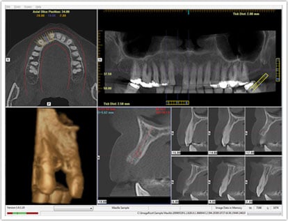
- Cone Beam Computed Tomography, or Cone Beam CT scanning, requires the patient to sit in the CT machine while the X-ray beam rotates around the head. A computer creates a three-dimensional image of the interior structures that is used to identify problems in the bones of the head and then to create treatment plans. Cone beam CT scanning is particularly useful in diagnosing and treating temporomandibular joint (TMJ) problems, which can be difficult to see with other types of X-rays.